Genomic Variants Explained: SNPs, Insertions, Deletions and Structural Variations
Explore genomic variation, from single nucleotide polymorphisms (SNPs) and small insertions or deletions to copy number changes and complex structural variants. Learn how genetic variants arise, how sequencing and bioinformatics identify them, and why their interpretation matters for genomics, evolution, disease research, and modern biotechnology.
Genomic Variants Explained: SNPs, Insertions, Deletions and Structural Variations
No two human genomes are completely identical. From differences in individual DNA bases to major chromosomal rearrangements, genomic variation helps explain biological diversity, evolution, inherited characteristics and the molecular basis of many diseases.
What Is Genomic Variation, and Why Does It Exist?
DNA stores hereditary information in a sequence of nucleotides: adenine, cytosine, guanine and thymine. Although individuals of the same species share much of their genetic information, their genomes contain numerous differences. These differences are collectively described as genetic or genomic variation.
Some variants are inherited from previous generations. Others arise through changes in DNA during replication, recombination or repair. Additional changes may occur in individual cells during an organism's lifetime. Those changes are especially important in the study of tissue mosaicism and cancer.
Variation is central to evolution because heritable differences create the genetic diversity on which natural selection and genetic drift act. Many variants have little or no detectable effect on phenotype, while others alter gene regulation, protein activity, chromosome organization or cellular survival.
A variant is not automatically harmful. It may be neutral, beneficial in a particular environment, associated with a measurable trait, or capable of contributing to disease. Interpretation therefore requires information beyond the existence of a DNA difference.
Single Nucleotide Variants and SNPs Explained
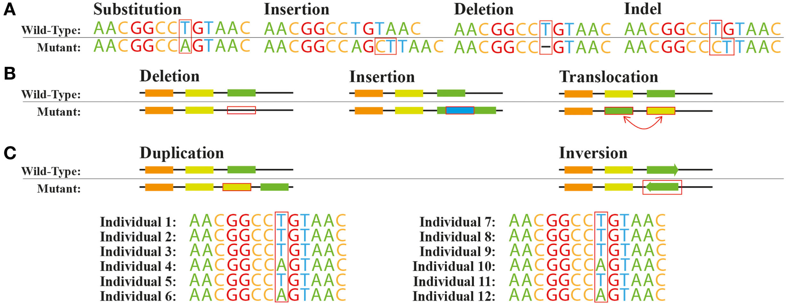
A single nucleotide variant (SNV) occurs when one DNA base differs from the corresponding base in a reference sequence. For example, a cytosine at a particular position may be replaced by thymine.
The term single nucleotide polymorphism (SNP) is commonly used for a single-base variant found at an appreciable frequency in a population, often operationally defined as at least 1%. SNV is the broader term and can describe both rare and common single-base differences.
A single-base variant may occur within a protein-coding exon, an intron, a regulatory sequence or an intergenic region. Within coding regions, some substitutions are synonymous: the encoded amino acid remains unchanged. Others are missense variants that change an amino acid, or nonsense variants that introduce a premature stop codon.
However, synonymous does not necessarily mean functionally irrelevant. Some synonymous changes can influence RNA splicing, transcript stability or translation. Similarly, a variant outside a protein-coding region may affect a promoter, enhancer or another functional regulatory element.
SNPs are widely used as markers in population genetics and genome-wide association studies. A statistical association between a SNP and a trait does not necessarily mean the SNP itself causes that trait; it may track another nearby causal variant through linkage disequilibrium.
A C G T A C G T
The sequence used for comparison.
A C G C A C G T
The fourth nucleotide differs: T → C.
Insertions and Deletions: How Indels Change DNA
An insertion adds one or more nucleotides relative to the reference sequence, whereas a deletion removes one or more nucleotides. Together, these changes are often called indels. The term can describe relatively small sequence changes, although large insertions and deletions may also be classified as structural variants.
The effect of an indel depends strongly on its length and location. Within a protein-coding sequence, an insertion or deletion whose length is not divisible by three usually shifts the reading frame. This changes the grouping of nucleotides into codons and may introduce a premature stop codon.
An indel whose length is divisible by three is generally called an in-frame insertion or deletion. It adds or removes amino acids without changing the reading frame downstream, but it can still interfere with protein folding, binding or enzyme activity.
Outside coding sequences, indels may influence transcription factor binding, RNA splicing or the architecture of regulatory DNA. Other indels may have no measurable functional consequence.
Accurate indel detection presents computational challenges because repetitive sequences can produce multiple plausible alignments. In such regions, the same biological event may initially be represented in different ways by variant-calling tools.
| Variant | DNA change | Potential consequence |
|---|---|---|
| SNV | One nucleotide substituted | Can change a codon, splice signal or regulatory element |
| Small insertion | One or more bases added | May create an in-frame change or a frameshift |
| Small deletion | One or more bases removed | May remove amino acids or change the reading frame |
| Complex small variant | Adjacent substitutions and indels | May require joint interpretation as one sequence event |
Structural Variants: Deletions, Duplications, Inversions and Translocations
Structural variants are larger changes in genomic organization. A commonly used operational definition includes events affecting at least approximately 50 base pairs, although precise conventions vary among studies and variant-detection methods.
Deletions remove genomic segments, potentially eliminating exons, complete genes or regulatory elements. Duplications create additional copies of DNA, while insertions introduce sequence at a new genomic position.
Inversions reverse the orientation of a genomic segment. Their effects depend on breakpoint positions and whether genes or regulatory relationships are disrupted. Translocations involve the relocation or exchange of DNA between chromosomal locations, often between different chromosomes.
Some structural variants change the amount of DNA present, while others rearrange the existing sequence without changing its overall copy number. These are often described as unbalanced and balanced structural changes, respectively.
Complex rearrangements may contain several event types together. Their interpretation can require knowledge of breakpoint sequences, chromosome context and the arrangement of multiple linked changes.

Structural variation is a significant source of genomic diversity because one event can affect far more nucleotides than a single-base substitution. This does not mean every structural variant is biologically disruptive; many are compatible with normal development and function.
Copy Number Variations and Their Biological Effects
Copy number variations, or CNVs, are differences in the number of copies of a genomic segment compared with a reference or another genome. Many CNVs arise through deletions and duplications.
In a typical diploid autosomal region, an individual usually carries two copies of a sequence, one inherited from each parent. A deletion may reduce this number, while duplication may increase it. Interpretation is more complicated in sex chromosomes, repeated sequences, mosaic tissues and polyploid organisms.
Copy number changes can influence gene dosage. Additional copies of a gene may alter the amount of RNA or protein produced, although regulatory feedback and other biological mechanisms can modify the relationship.
CNVs can also disrupt genes at their boundaries, change enhancer-promoter relationships or alter chromatin organization. Their functional effects depend on the affected genomic content rather than copy number alone.
Researchers detect CNVs using approaches such as sequencing read-depth analysis, SNP arrays, comparative genomic hybridization and other genomic methods. The most appropriate method depends on event size, sample characteristics, resolution requirements and research objectives.
| Structural change | Usually alters DNA copy number? | Possible functional effect |
|---|---|---|
| Deletion | Yes, decreases it | Loss of coding or regulatory material |
| Duplication | Yes, increases it | Altered gene dosage or regulatory organization |
| Inversion | Not necessarily | Gene disruption at breakpoints or altered regulation |
| Balanced translocation | Not necessarily | Breakpoint effects, altered regulation or gene fusion |
Germline, Somatic and Mosaic Genetic Variants
Genomic variants can also be classified by their biological origin. Germline variants are present in reproductive cell lineages and may be transmitted to offspring. An inherited germline variant can be present in many or all tissues of an individual.
Somatic variants arise in cells outside the reproductive lineage during development or later life. They can result from DNA replication errors, environmental exposures or changes in DNA maintenance mechanisms.
Somatic variation is especially important in cancer genomics. Tumors accumulate genetic changes, some of which contribute to abnormal cell growth or survival. Many others are passenger changes that do not directly drive tumor development.
Mosaicism occurs when genetically distinct cell populations coexist within an organism. A variant arising early in development may be distributed among multiple tissues, whereas a later change may remain restricted to a smaller cell population.
Differentiating germline from somatic variants can require comparison with appropriate non-tumor samples, analysis of allele fractions and consideration of biological context. Allele fraction alone is not always sufficient to establish the origin of a variant.
| Variant category | Origin | Common research context |
|---|---|---|
| Germline | Inherited or arising in a reproductive lineage | Inherited traits, population genomics, hereditary disorders |
| Somatic | Arises in a non-reproductive cellular lineage | Cancer genomics, aging, tissue-specific mutations |
| Mosaic | Variant present in only a subset of cells | Developmental biology, tissue heterogeneity, mosaic disorders |
How DNA Sequencing and Bioinformatics Detect Variants
Modern genomic analysis often begins by sequencing DNA from a biological sample. The resulting reads contain information about nucleotide sequences from many genomic positions, but the raw data cannot immediately be interpreted as a reliable list of variants.
Researchers first evaluate read quality, sequencing artifacts and sample characteristics. In many conventional workflows, reads are aligned to a reference genome so that differences between the sample and reference can be examined.
Variant calling applies computational methods to identify supported sequence differences. The analysis considers evidence such as base quality, alignment quality, read depth, local sequence context and the balance of reads supporting different alleles.
Different tools are optimized for different variant classes. A method that detects SNVs and small indels reliably may not detect a complex inversion or large insertion with comparable sensitivity. Structural variant callers often analyze additional signals such as split reads, discordant read pairs, coverage changes or assembled sequence.
After calling, researchers normalize variant representations, apply appropriate quality filters and annotate genomic positions. The final variant set must be interpreted in the context of research aims and dataset limitations.

An important practical lesson is that variant detection depends on the whole analytical workflow. Sequencing quality, read length, reference selection, alignment strategy and variant caller can all influence the results.
Short-Read, Long-Read and Whole-Genome Variant Analysis
The choice of sequencing technology influences which genomic variants can be detected. Short-read sequencing has become a widely used approach for identifying SNVs and small indels in accessible genomic regions.
Short reads can struggle to resolve large insertions, repeated sequences and complicated rearrangements because individual reads may not span the relevant genomic structure. Paired-end information and specialized algorithms improve detection, but some ambiguities remain.
Long-read sequencing provides reads capable of spanning larger structural events. This can help identify insertion sequences, determine rearrangement breakpoints, resolve repeated regions and phase linked variants onto individual haplotypes.
Whole-exome sequencing (WES) focuses primarily on captured protein-coding exons. It can be useful for questions centered on coding variation but does not provide comprehensive coverage of the entire genome.
Whole-genome sequencing (WGS) surveys both coding and noncoding regions. Its greater genomic breadth supports investigation of more diverse variant classes, although detection performance still depends on sequencing technology, coverage and analysis.
| Approach | Typical strength | Important limitation |
|---|---|---|
| Short-read WES | SNVs and small indels in well-covered targeted coding regions | Limited access to most noncoding regions and many structural events |
| Short-read WGS | Broad genomic assessment of small variants and some SVs | Repeat-rich and complex regions may be difficult to resolve |
| Long-read WGS | Structural variants, complex regions and haplotype phasing | Cost, input requirements and error profiles vary across implementations |
| Complementary genomic methods | Targeted confirmation and copy-number or chromosome-level analysis | Usually optimized for particular classes and scales of variation |
Increasingly, genomics studies integrate multiple data types. A combination of short-read, long-read and complementary methods may provide a more complete representation of structural variation than any one strategy alone.
Variant Annotation, Population Databases and Interpretation
Once a variant has been detected, an important question remains: what does it mean? Variant annotation connects sequence differences to genomic features such as genes, transcripts, exons, regulatory regions, known population alleles and previously documented observations.
Functional annotation may classify coding consequences as synonymous, missense, frameshift or stop-gain changes. For regulatory and noncoding variants, interpretation often relies on additional molecular evidence such as expression studies, chromatin accessibility or experimentally characterized regulatory elements.
Population resources help determine whether a variant is frequently observed in particular populations or appears exceptionally rare. Resources such as gnomAD support population frequency assessment. dbSNP catalogs many small variants, while ClinVar provides submitted interpretations concerning relationships between genetic variants and human conditions.
Database inclusion does not, by itself, demonstrate pathogenicity. Similarly, a rare variant is not necessarily harmful, and a common variant may still influence a complex trait. Reliable interpretation requires multiple lines of evidence.
A further challenge arises from reference bias. Most routine workflows compare reads against a linear reference genome. Regions that diverge substantially from that reference may be inaccurately represented, misaligned or missed.
Pangenome references offer a broader way to represent genetic diversity by including multiple alternative genomic paths. This approach is particularly important for complex regions and structural variants.

How Variant Analysis Advances Biotechnology and Scientific Discovery
The analysis of genomic variants provides a connection between DNA sequence and biological diversity. In population genomics, variant patterns help researchers study ancestry, demographic history, adaptation and evolutionary change.
In disease research, genetic variants can help identify genes and biological pathways associated with particular conditions. The interpretation of rare variants often requires family information, functional evidence and knowledge of established inheritance mechanisms.
Cancer genomics investigates somatic substitutions, insertions, deletions, copy-number alterations and structural rearrangements. Their combined patterns can reveal aspects of tumor evolution and molecular mechanisms, while distinguishing drivers from passengers remains a central research challenge.
In microbial and agricultural biotechnology, variant analysis can support investigations of strain diversity, evolutionary adaptation, metabolic traits, pathogen populations and genetic changes introduced through engineering.
Emerging technologies are extending genomic discovery beyond small variants. Long-read sequencing, improved genome assemblies, haplotype phasing and pangenome graphs help reveal variation in regions that were previously difficult to analyze.
At the same time, integrating genomic variation with NGS data analysis, transcriptomics, proteomics and computational biology allows researchers to explore how sequence differences influence molecular systems rather than examining DNA changes in isolation.
The long-term scientific goal is not only to catalog genomic differences, but to understand when they matter, through which mechanisms, and under which environmental and biological conditions. This transformation from sequence data to functional knowledge is a defining challenge of modern genomics.